Bead Mill Homogenization Streamlines Dermatomycoses Organism Identification by Drastically Reducing Sample Preparation Time
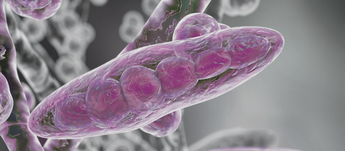
Pathogenic fungi, i.e. dermatophytes, are responsible for superficial infections known as dermatophytosis, which occurs in humans and animals (1, 2). These infections occur in nails, skin, and hair because of the high- affinity dermatophytes have for keratin and keratin-like structures (2-4). In addition to dermatophytes, non- dermatophyte molds and yeast can cause dermatomycosis. The pathogenesis of dermatophytosis involves secreting multiple enzymes, such as serine-subtilisins and metallo-endoproteases (fungalysins), formerly called keratinases. Enzymes such as hydrolases, lipases, and ceramidase are mucolytic enzymes that help penetrate keratin-rich tissue, providing nutrition to the fungi (2, 3). Human fungal infection is growing, creating a worldwide epidemic (4, 5). These fungi rarely cause infections in healthy individuals; however, their presence can be severe for individuals with depleted or compromised immune systems, such as those with HIV/AIDS, hence the importance of proper fungi identification and treatment (6, 7). Advances in laboratory technology offer time and cost savings; however, accurately identifying the presence of dermatophytes in humans and animals remains challenging.
Currently, the identification of dermatophytes and other pathogenic organisms is determined by mycological testing using culture, microscopic, and molecular techniques, which are time-consuming, not specific, or limited to the number of detectable fungi (9, 10). For example, one of the major complications associated with the macroscopic examination of fungi is the subjectivity of the morphological evaluation (4, 11). Likewise, microscopic examination of the structures found on fungi grown in culture may also be challenging to determine since morphology has been shown to change over time and may require laboratory personnel with extensive experience and expertise (11). In addition, culture has yielded negative results, even after visual examination confirmed the presence of arthrospores, suggesting possible isolation from non-dermatophyte fungi (Dermatomycosis) (4, 11). Molecular techniques such as traditional or real-time PCR have shown to be very selective, allowing accurate identification of fungi; however, the number of species that can be identified using the same primer set is limited and does not cover the full dermatophyte spectrum (4, 11). Therefore, species-specific identification is critical.
The number of dermatophytes identified continues to increase, partly due to advancing molecular techniques (12). The EUROArray Dermatomycosis system is a highly sensitive and specific molecular platform for identifying 50 dermatophyte species, 3 yeasts, and 3 molds. However, the time spent during sample preparation, specifically overnight digestion of nail samples, is the current lynchpin. Here we show a novel approach to sample preparation for identifying dermatophytes and non-dermatophyte origin. Our method included homogenization to disrupt the nail sample and any fungi present, producing a uniform homogenate. Nucleic acids of host and pathogenic origin are released, quantified, and identified as dermatophyte or non- dermatophyte using the EUROArray Dermatomycosis system for molecular genetic direct identification of the presence of dermatophytes, yeasts, and molds. Using homogenization, we significantly reduced the required sample preparation time thus improving sample throughput.
